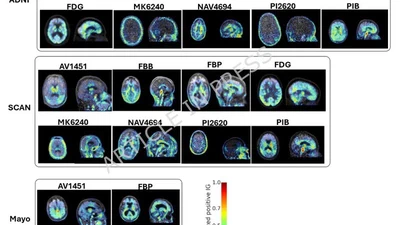
Automated PET tracer classification in multi-site, multi-cohort studies using deep learning
Deep learning classifies brain PET radiotracers directly from scans across sites and cohorts, improving metadata quality control.
robel-k.-gebre
 RG
RGI use deep learning, explainable AI, and disease progression models to turn medical images into useful measures of disease. My current work focuses on tau PET quantification, MRI harmonization across sites, and imaging differences between multiple system atrophy and Parkinson’s disease.
I created the THETA score, a tau PET measure that captures the spatial pattern of tau across the brain.
My publications also appear under Robel K. Gebre and Robel Kebede Gebre.
Deep learning classifies brain PET radiotracers directly from scans across sites and cohorts, improving metadata quality control.
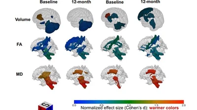
We developed a machine learning framework to quantify disease heterogeneity and progression in multiple system atrophy and Parkinson's disease using structural and diffusion MRI.
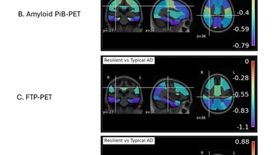
We applied the THETA score to investigate cognitive resilience mechanisms in individuals with heterogeneous global tau burden.
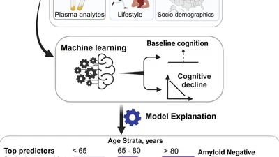
MRI, plasma biomarkers, genetics, cardiovascular risk, and lifestyle measures were compared for predicting cognitive decline.
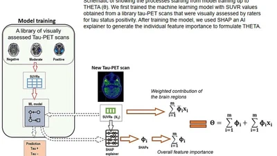
THETA is a machine-learning tau PET measure that captures the spatial pattern of tau deposition across the brain in Alzheimer's disease.
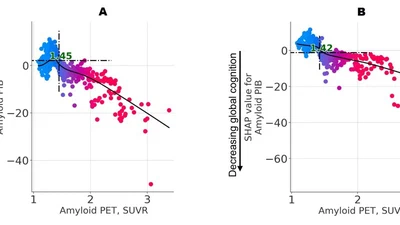
We developed a machine learning method to identify transition points in Alzheimer's disease biomarkers as a supplement to the reliable worsening method.
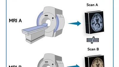
A systematic comparison questions the reliability of widely used cross-scanner harmonization methods for structural MRI.
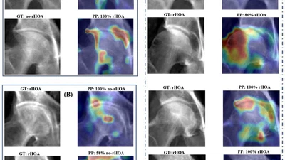
We developed the first deep learning application for detecting hip osteoarthritis from 2-D summation images derived from routine clinical CT scans.

I will remotely attend AAIC 2026 in London to present recent work in neuroimaging and machine learning for Alzheimer's disease.


A podium talk on a 2.5D deep learning classifier for PET tracer identification, and a poster introducing the tau therapeutic window as an eligibility criterion for anti-amyloid …
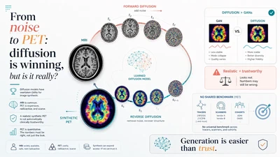
Diffusion methods have overtaken GANs for generating images. Turning MRI into PET is within reach, yet a realistic picture is not a trustworthy one.
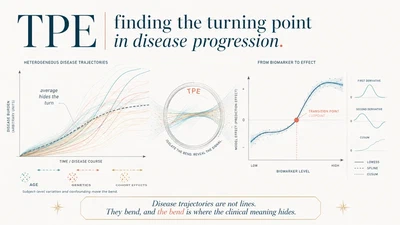
Disease trajectories are not lines. They bend, and the bend is where the clinical meaning hides.
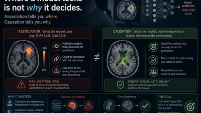
Explainability tools tell you which features a model used. They rarely tell you which ones mattered.
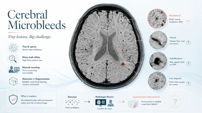
Why cerebral microbleeds are difficult to detect and segment on MRI, and what they mean for small vessel disease and anti-amyloid therapy safety.